Diagnosis & Imaging
Getting to the diagnosis, then confirming it. This section brings together ECG interpretation, bedside echocardiography, cross-sectional imaging and structured differential diagnosis for the presentations that cause most difficulty: undifferentiated chest pain, unexplained breathlessness, syncope, an abnormal tracing with a well-looking patient. It is meant for clinicians who read their own studies because no reporting cardiologist is available the same day. Commonly used are Clinical ECG Interpretation, Cardiac Imaging: CT and MRI and the 1,800-case diagnosis reference, which together cover most of what walks through a general clinic.
Investigations that change managementDiagnosis and imaging titles are organised around what a test will alter rather than what it can display. Echocardiography is covered at reporting level with normal values and the measurements that are commonly mis-taken; cross-sectional imaging is covered by the question it answers, so CT, cardiac MRI and nuclear studies are placed against the clinical problem rather than described as modalities. Considerable space is given to the negative decision — which murmur does not need serial imaging, which chest pain does not need an angiogram, which incidental finding needs nothing at all. Overinvestigation is the commonest error in general cardiology and the hardest to unlearn, and these titles address it directly.
Showing all 6 results
-
Sale!

AI in Cardiology — Diagnosis, Care and Management
Original price was: ₹4,100.00.₹1,499.00Current price is: ₹1,499.00. Add to cart -
Sale!
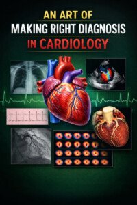
Bedside Cardiology Diagnosis — Getting It Right
Original price was: ₹6,400.00.₹1,450.00Current price is: ₹1,450.00. Add to cart -
Sale!
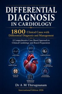
Differential Diagnosis in Cardiology — 1,800 Cases
Original price was: ₹7,400.00.₹3,070.00Current price is: ₹3,070.00. Add to cart -
Sale!
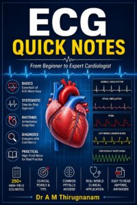
ECG Quick Notes — Clinical Reference
Original price was: ₹3,300.00.₹1,450.00Current price is: ₹1,450.00. Add to cart -
Sale!
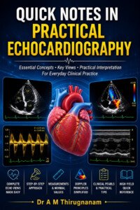
Echocardiography Quick Notes — Practical Reference
Original price was: ₹4,100.00.₹1,760.00Current price is: ₹1,760.00. Add to cart -
Sale!
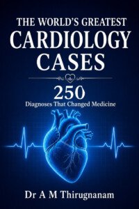
The World’s Greatest Cardiology Cases — 250 Diagnoses
Original price was: ₹3,200.00.₹1,450.00Current price is: ₹1,450.00. Add to cart